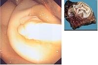
Hydatid cyst (pulmonary echinococcosis)
-Whitish-yellow, bright, laminated, gelatinous membranes of a hydatid cyst visualised during bronchoscopy.
-The bronchoscope is in the interior of the hydatid cyst. The latter communicated with a bronchus.
-The inset image shows a cross section through the lung and the hydatid cyst.
Educational Material:
-Endobronchial findings of hydatid cyst disease: a report of five pediatric cases. Kut A et al. Pediatr Pulmonol 2012;47(7):706-9.
-Bronchoscopic diagnosis and removal of a ruptured hydatid cyst. Sharif A et al. J Bronchology Interv Pulmonol 2011;18(4):362-4.
-Spontaneous Expulsion of a Hydatid Cyst During Flexible Bronchoscopy. Singh B et al. J Bronchology 2006;13(4):214-5.
- Bronchoscopic diagnosis of a ruptured hydatid cyst in a young male with hemoptysis. Koul PA et al. J Bronchology Interv Pulmonol 2011;18(1):61-4
- Bronchoscopic Diagnosis of Pulmonary Hydatidosis in Patients With Unusual Roentgenologic Appearance. Gupta D et al. J Bronchology 2001;8(2):101-3
- Intrabronchial tumor: An atypical presentation of a hydatid cyst. Gamez BJ et al. J Bronchology 2003;10(3):192-4

